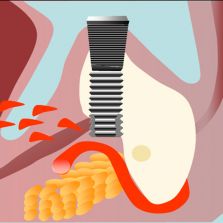
CBCT - Implantology planning
- the width and height of bone available - the best angle at which to set the implant - the presence of bone undercuts - the rapport between compact bone and bone marrow - the presence of diseases - the exact localisation of anatomical structures such as maxillary sinus, the lower alveolar canal and the mental foramen. Primary x-rays, endoral and a panoramic x-rays are not sufficient to obtain all this information as their factors of enlargement and deformation are difficult to calculate and especially as they do not allow the viewing of cross sections that a surgeon needs to identify individual anatomical structures nor the relationship they have with adjoining structures. To be able to obtain this information, a dentist has to therefore avail of tomography. Continues on the book: Atlas of Cone Beam - Volumetric 3D Images - Edizioni BDD.
|


